A panoramic X-ray, also known as an orthopantomogram, is a widely used diagnostic imaging method in dentistry. It provides a comprehensive view of the upper and lower jaws, teeth, joints, and surrounding structures, helping the dentist accurately assess oral health.
It allows the detection of issues such as cavities, periodontal diseases, cysts, tumors, and other abnormalities that may not be visible to the naked eye or with other diagnostic methods.
It aids in the planning of complex dental treatments, such as implants, tooth extractions, orthodontic treatments, and restorations. The panoramic image provides a clear view of the position and condition of the teeth and jawbones.
It is used to monitor the progress and effectiveness of dental treatments, allowing the dentist to evaluate progress and adjust treatment if necessary.
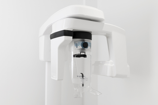
The patient is positioned in a special place in front of the orthopantomograph and is asked to bite on a plastic support to stabilize the head and lower jaw.
The digital orthopantomograph rotates around the patient’s head, taking a panoramic image of the oral cavity. The entire process takes a few seconds.
The digital image appears immediately on the computer screen, allowing the dentist to evaluate it and identify any problems.
In our clinic, we use the CARESTREAM digital orthopantomograph, which offers high-resolution images with minimal radiation. Digital technology ensures quick and accurate diagnosis, enabling the dentist to detect even the smallest details and plan the best treatment for the patient.