
Herpes Labialis
Herpes labialis is caused by the herpes simplex virus type 1 (HSV-1), and much more rarely by HSV-2. It manifests with the characteristic blisters in the perioral area. These blisters are small, fragile, and often clustered together in one spot.
Symptoms of herpes labialis include pain, burning, and a tingling sensation, which appear before the blisters erupt.
The frequency of herpes labialis or cold sores varies from person to person. It can appear many times a year or be limited to 1 or 2 episodes in a lifetime. Usually, the number of episodes of cold sores decreases over time.
Cold sores are highly contagious. They are typically transmitted through saliva and direct contact. For most patients, no specific treatment is required.
Since the virus cannot be permanently removed from the body, the treatment aims to manage the lesions.

Aphthous Ulcers
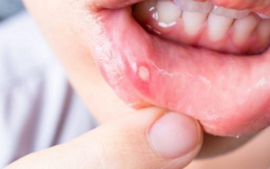
Aphthous ulcers are sores that appear on the oral mucosa, which is lined with non-keratinized epithelium, e.g., the tongue, cheeks, etc.
They are accompanied by intense pain and often have periods of flare-ups and remissions that can range from a few days to several months or even years. They are classified as minor (less than 10 mm in diameter), major (greater than 10 mm in diameter), and herpetiform ulcers, which are multiple clustered aphthous ulcers.
Aphthous ulcers are benign, self-healing lesions, meaning they will heal on their own within a reasonable period. The exact cause is unknown, so treatment focuses on patient relief. Treatment may include topical medications in the form of mouth rinses, sprays, or gels, as well as systemic medications taken orally, such as corticosteroids, anti-inflammatories, and antimicrobials.

Leukoplakia
Leukoplakia is a potentially malignant lesion that appears on the oral mucosa in the form of a white plaque, with an increased likelihood of developing into squamous cell carcinoma.
Regular alcohol consumption, neglect of oral hygiene measures, and especially smoking are the main factors leading to the appearance of leukoplakia.
It is an asymptomatic lesion and there are three different types: homogeneous leukoplakia, which is the most common (97% of cases) and has the lowest rates of cancer development; granular leukoplakia, which presents the highest risk of malignant transformation; and verrucous leukoplakia, which also presents a significant risk of malignant transformation.
Treatment of leukoplakia involves the surgical excision of the lesion. The patient must quit smoking, otherwise, the risk of recurrence or malignant transformation of leukoplakia is high.
Equally important factors for ensuring the successful outcome of the treatment include maintaining oral hygiene, addressing potential mouth injuries, and regular patient monitoring (every 4 to 6 months).

Burning Mouth Syndrome
Burning mouth syndrome can occur in any area of the mouth, often covering a large extent. Patients may perceive and describe their symptoms as a “burning sensation in the mouth” or “tingling in the mouth,” often accompanied by “dryness” and a “strange or unpleasant taste.”
These symptoms often persist for several weeks, months, or even years without significant changes. The intensity of the burning mouth syndrome symptoms varies and can remain constant or fluctuate throughout the day.
Burning mouth syndrome can be idiopathic or secondary. In idiopathic cases, the burning pain appears in the oral cavity without any systemic pathological condition related to the disease. In secondary forms, burning mouth syndrome coexists with other diseases and conditions (e.g., vitamin deficiencies).
Treatment for burning mouth syndrome involves addressing the underlying cause and providing relief to the patient.