
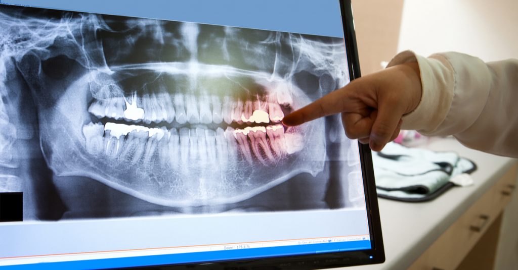
Radiography serves as a diagnostic tool, allowing the dentist to obtain information about early cavities between the teeth, cavities under existing restorations, root fractures, periapical lesions or cysts, periodontal disease, the presence of impacted teeth, and more. This information is essential for accurate diagnosis, treatment planning, monitoring therapy, and tracking the progression of a lesion.
As many dental issues can only be diagnosed radiographically, the dentist can use radiographs to detect oral cavity problems earlier. This early detection helps prevent severe pain, allows for the prompt treatment of the issue, and ensures a better prognosis for the therapeutic restoration.
Depending on the case, the dentist decides which type of radiograph is required. The most commonly used are:
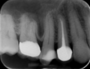
The periapical radiograph.
The panoramic radiograph.
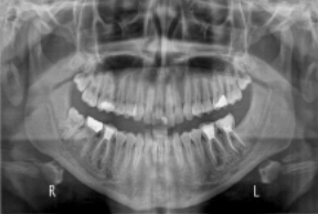
The panoramic radiograph has become the simplest radiographic examination that displays all the hard tissues. It allows the dentist to view all the teeth and the bone of the upper and lower jaw, as well as the temporomandibular joints, in a single image. It is primarily used for diagnosing dental, periodontal, and bone diseases, as well as for evaluating and planning treatments or surgical interventions.
Its drawback is that it lacks high resolution, so not all details are captured, and it provides a two-dimensional image of a three-dimensional object, such as the jaw and teeth.
The CT Scan.
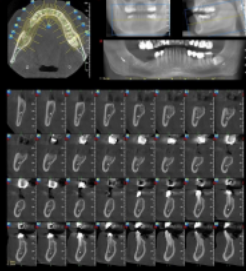
It is a specialized radiograph that provides high-quality and accurate three-dimensional imaging of the structures of the upper and lower jaw and the teeth. It is used for the study and planning of treatments involving implants, bone regeneration of jaw lesions with bone grafts, impacted teeth, periapical lesions not identified in panoramic radiographs, and more. Jaw CT scans have become the most modern and important diagnostic method in dentistry, gaining ground in clinical practice every day.
Suggested Reading:
Exercises in Radiodiagnosis for Intraoral Radiographs**, University Studio Press, Scientific Book and Journal Publications.
Dental Radiography**, Evangelos Pantelis, Assistant Professor of Medical Physics, Laboratory of Medical Physics, Medical School of Athens.